We provide specialist dental care in Oral & Maxillofacial Surgery and Orthodontics.
Dental Cyst
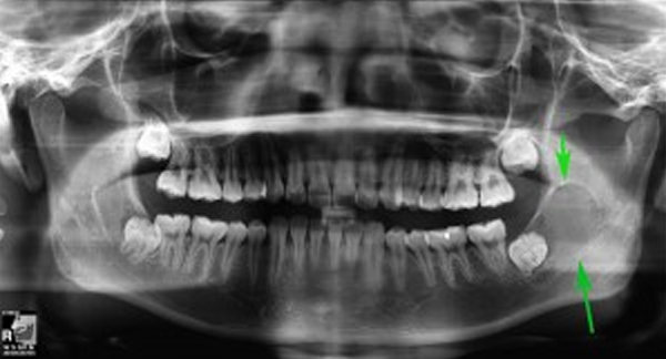
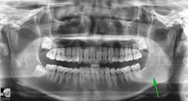
Dentigerous cyst, which is associated with buried tooth
Radicular cyst, which is associated with infection at the root tips of a tooth
Primordial cyst, which is formed from residual cells from developing tooth buds.
Keratocyst, a special category that has great propensity to recur following treatment. As such, this lesion has been re-classified as “Keratocystic Odontogenic Tumour”.
Unicystic ameloblastoma, an odontogenic tumour which is locally invasive.
Marsupialisation
The cyst is surgically “burst” opened. As the cystic membrane merges with the oral mucosa, the bone defect refills. A time interval of up to a year may be needed and only the benign cyst-type is suitable for this treatment modality.
Enucleation
The lining of the cyst is separated from the bone and surrounding tissues. The resulting bony defect may be treated with burring, cryotherapy, laser or chemicals such as Carnoy’s solution.
Resection
A segment of the bone is surgically removed along with the cyst. The discontinuity defect is reconstructed with bone graft harvested from another site such as the hip bone or leg (femur). Titanium bone plates and screws are used to fixate the bone.
Decompression
Occasionally, a large cyst is “marsupialised” first to allow it to shrink before being “enucleated”.